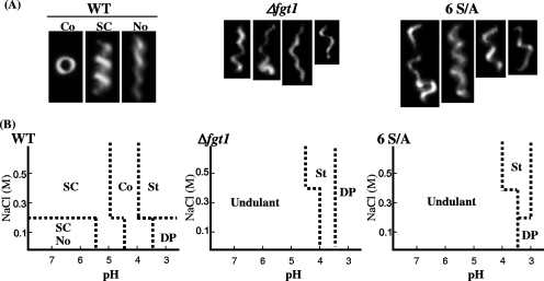
FIG. 3.
Polymorphic transitions of flagellar filaments from the WT and glycosylation-defective mutants of P. syringae pv. tabaci 6605. (A) Dark-field light micrographs of flagella. Typical images of Coiled, Semi-Coiled, and a mixture of Semi-Coiled and Normal filaments prepared from the WT and undulant filaments prepared from Δfgt1 and 6 S/A mutant strains. (B) Phase diagrams of polymorphs by pH and NaCl concentration. SC, Semi-Coiled; No, Normal; Co, Coiled; St, Straight; DP, depolymerized.

