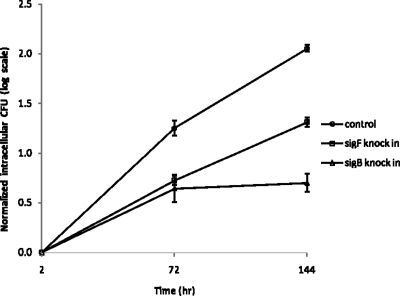
FIG. 4.
Intracellular growth of M. tuberculosis sigB and sigF KI strains in activated J774A.1 macrophages in vitro. Murine macrophage J774A.1 cells were activated with gamma interferon and lipopolysaccharide (as in Materials and Methods) and then infected with M. tuberculosis strains harboring the empty vector, the sigB KI vector, or the sigF KI vector at a multiplicity of infection of 1:1. After incubation for 2 h, the macrophage monolayer was extensively washed, and samples were taken to determine the initial intracellular CFU titer. The bacterial growth rate was determined by CFU counting of bacilli at 3 and 6 days after infection. The log CFU counts are normalized to the initial intracellular CFU counts.

