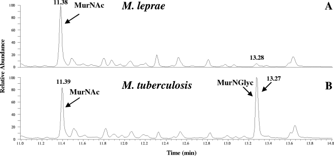
FIG. 2.
Analysis of the muramic acid residues of PGs from M. tuberculosis and M. leprae. Shown are the total-ion chromatograms for M. leprae and M. tuberculosis. The trimethylsilane derivatives of the muramic acids were analyzed by GC-MS as described in Materials and Methods. The peak at 13.28 min in panel A does not correspond to MurNGlyc, as determined by MS.

