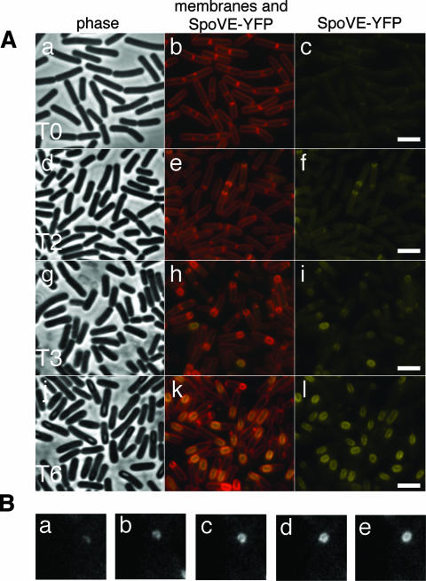
FIG. 1.
SpoVE-YFP localization throughout sporulation. (A) Strain AH3561 (spoVE85 amyE::spoVE-yfp) was induced to sporulate in DSM medium, and samples were collected at the onset (0 h [T0]) and throughout sporulation and labeled with vital stain FM4-64 for visualization of membranes by fluorescence microscopy. The first column shows phase contrast images and the second column depicts the colocalization of the membranes (red) and the YFP signal (yellow). The third column shows the localization of SpoVE-YFP alone (yellow). Scale bars (white) represent 1 μm. (B) Strain JDB1813 carrying an SpoVE-GFP fusion was visualized by time-lapse microscopy every 7 min as it proceeded through sporulation. Shown is a single cell with images taken at five consecutive time points (a, t = 0; b, t = 7 min; c, t = 14 min; d, t = 21 min; e, t = 28 min).

