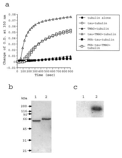
Figure 1.
(a) Spectrophotometric measurement of tubulin assembly. Tubulin was incubated (30°C, 15 min) alone (solid circles) or with TMAO (solid triangles), and the assemblies were monitored by an increase in the absorbance at 350 nm. By using the same conditions, tubulin was assembled in the presence of tau (open circles) or tau and TMAO (open triangles). Finally, tubulin was assembled with PKA-phosphorylated tau in the presence (open squares) or absence (solid squares) of TMAO. The phosphorylation stoichometry of PKA-phosphorylated tau was 2.7 ± 0.1 phosphates/mol. (b) SDS/PAGE analysis of tau proteins. Nonphosphorylated tau (lane 1) and PKA-phosphorylated tau with 2.8 phosphates/mol (lane 2) were analyzed in a 10% SDS/PAGE gel. Proteins in the gel were visualized by a Coomassie brilliant blue R staining. (c) Radioautogram of 32P-labeled tau in a SDS/PAGE analysis. Tau was phosphorylated with PKA in the presence of [γ-32P]ATP. Any aggregates of phosphorylated tau–TMAO (lane 1) and phosphorylated tau—tubulin–TMAO (lane 2) were collected by centrifugation and analyzed by SDS/PAGE.

