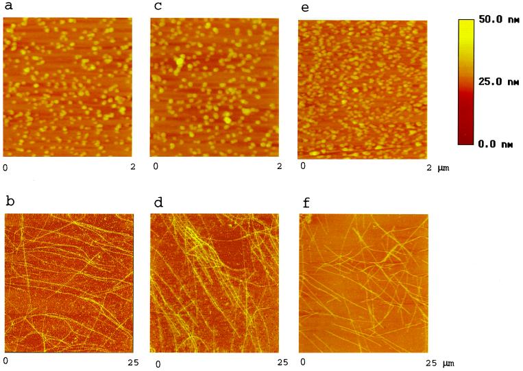
Figure 2.
AFM images of tubulin assembly. Tubulin (a) was assembled by using standard reaction conditions (Fig. 1) and fixed. The mixture was then spotted onto a mica disc, dried, and scanned by AFM. Other reaction mixtures that included tau (b), TMAO (c), tau + TMAO (d), PKA-phosphorylated tau with 2.7 phosphates/mol (e), or PKA-phosphorylated tau + TMAO (f) were also scanned. The sample height is color coded, as indicated by the height scale bar. Note field sizes for a, c, and e (2 μm × 2 μm) are smaller than those for b, d, and f (25 μm × 25 μm) to highlight features. The heights of MTs shown in b, d, and f are all about 20 nm, in general.

