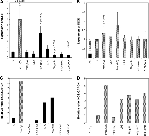
FIG. 5.
Endothelial expression of iNOS also results from TLR ligand challenge. Nonactivated or activated cells were incubated with TLR-specific ligands for 24 h, and a two-step quantitative real-time RT-PCR (A and B) or a two-step RT-PCR and determination of band intensity (C and D) were performed to detect the expression of iNOS relative to those of 18S mRNA and GAPDH, respectively. Black bar, absence of cytokines; hatched bar, presence of cytokines; column C, control; column C + Cyt., control in the presence of cytokines; columnn C − Cyt., control in the absence of cytokines. (A) In control HMEC-1, borderline expression of iNOS is found (CT = 33.5). Only the addition of poly(I:C) results in the de novo synthesis of iNOS. Upon challenge with flagellin or CpG DNA, the basal expression of iNOS is significantly reduced. (B) When activated with a triplet of proinflammatory cytokines (IL-1β, TNF-α, and IFN-γ; 1,000 U/ml each [C + Cyt]), iNOS expression is significantly increased as expected, and the addition of specific ligands results in a further increase in iNOS expression only with Pam3Cys. (C) In control HDMEC-1, iNOS is not expressed, but the addition of LPS and flagellin results in the de novo synthesis of iNOS mRNA. (D) With activated primary cells (as described for panel B), de novo synthesis of iNOS is observed. An additional increase in iNOS expression is seen with all ligands except poly(I:C) (n = 1 to 4).

