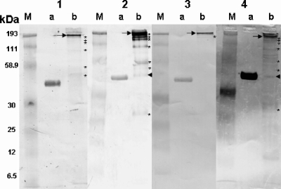
FIG. 3.
Western blot analyses of transferred proteins on nylon membranes. The antibodies in the serum from a human with lacaziosis weakly detected Paracoccidioides brasiliensis gp43 (lane 1a). This serum strongly detected an ∼193-kDa immunodominant antigen (arrow) as well as six other weak bands (asterisks) (lane 1b). The IgG in the serum from the experimentally infected mouse weakly detected P. brasiliensis gp43 (lane 2a). The mouse antibodies strongly detected the ∼193-kDa immunodominant antigen (arrow) as well as seven other proteins (asterisks) and an ∼48-kDa protein not detected by the other sera (lane 2b, arrowhead). The antibodies in the dolphin with lacaziosis slightly detected P. brasiliensis gp43 (lane 3a). The antibodies strongly recognized the ∼193-kDa immunodominant antigen (arrow) and only one of the minor antigens previously detected in the L. loboi extract (lane 3b). The antibodies in the serum from the patient with paracoccidioidomycosis strongly detected gp43 (lane 4a). The antibodies of this patient also detected the immunodominant ∼193-kDa antigen (arrow) in the L. loboi extract as well as other antigens (asterisks), including a 43-kDa antigen (arrowhead) (lane 4b). The molecular mass marker (lanes M) appears in each of the panels.

