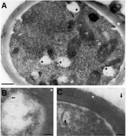
FIG. 5.
TEM of C. neoformans suggesting the presence of cytoplasmic vacuole-containing vesicles reminiscent of exosome-like structures. (A) Overview of a C. neoformans cell with different cytoplasmic vacuoles containing vesicles (black asterisks). The white asterisk indicates the cell wall. Scale bar, 500 nm. A magnified view of the vesicle-containing vacuoles is shown. Panel B demonstrates that these structures are surrounded by a bilayered membrane, which sometimes invaginates (arrow). A close association with the cell wall (white asterisk) was observed, suggesting fusion with the plasma membrane. Scale bar, 200 nm. (C) Intracellular and extracellular vesicles (black arrows) have similar dimensions. Scale bar, 200 nm.

