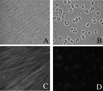
FIG. 3.
Effect of mycalolide B on HFFs. HFFs were exposed for 5 min either to solvent alone in medium (control) or to 3 μM mycalolide B. They were then washed, and 48 h later, they were photographed. (A) Phase micrograph of control HFFs. (B) Phase micrograph of HFFs exposed to mycalolide B. (C) Phalloidin staining of cells in panel A, showing microfilaments. (D) Phalloidin staining of cells in panel B, showing continued disruption of microfilaments.

