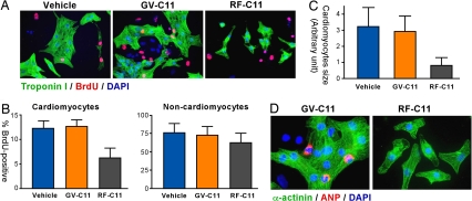
Fig. 6.
RF-C11 inhibits the proliferation and hypertrophy of cardiomyocytes. (A) Cells from mouse embryonic hearts were treated with BrdU for 12 h in the presence of 25 μM compound (RF-C11 or GV-C11) or solvent alone (vehicle; 0.25% EtOH in PBS), followed by immunostaining troponin I (for cardiomyocytes, green) and BrdU (for S-phase cells, red). (B) BrdU indices were determined by counting BrdU-positive cells for cardiomyocytes (troponin I-positive) and noncardiomyocytes (troponin I-negative). At least 400 cells per group were counted for the BrdU incorporation assay. (B and C) Data represent mean ± SEM obtained from three independent experiments. (C) The surface areas of troponin I-positive cells were determined by using >1,000 cells per group by using ImageJ software version 1.34s (National Institutes of Health). (D) Cardiac cells treated with RF-C11 or GV-C11 were immunostained for sarcomeric α-actinin (for cardiomyocytes, green) and ANP (for hypertrophism, red) with DAPI counterstaining.

