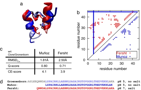
Fig. 2.
Comparisons of independently determined NMR structures for variant protein constructs of BBL. (a) Superimposed ribbon diagrams of the NMR structures of BBL as resolved by the groups of Clore and Gronenborn (light blue), Muñoz (blue), and Fersht (red). (b) Contact map of the Muñoz (blue) and Fersht (red) structures with a distance cutoff of 9.0 Å. The solid blue lines indicate where the Muñoz structure was truncated. (c) Table of the Cα-RMSD, Q-score, and CE-scores of the Muñoz and Fersht structures with respect to the Clore/Gronenborn structure. (d) Sequences of the structured residues in the NMR structures of the protein constructs obtained by the groups of Clore and Gronenborn (light blue, structured; gray, unstructured), Muñoz (blue), and Fersht (red).

