
Fig. 3.
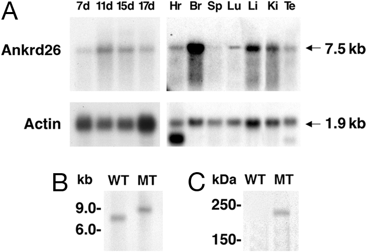
Expression analysis of Ankrd26 gene. (A) Northern blot showing the expression of Ankrd26 transcript in various tissues. The RNA samples used in the analysis are from heart (Hr), brain (Br), spleen (Sp), lung (Lu), liver (Li), kidney (Ki), testis (Te), and total embryos from 7 days (7d), 11 days (11d), 15 days (15d), and 17 days (17d). The blots were probed with actin gene to show the quality as well as equal loading of RNA in each lane. (B) Northern analysis showing the inactivation of the Ankrd26 transcript in mutant mice. About 20 μg of total RNA from brain of WT and MT mice was analyzed by Northern blot hybridization as described in Materials and Methods. (C) Detection of Ankrd-lacZ fusion protein in MT mice. Tissue extracts from MT and WT liver were analyzed by Western blotting with anti-β-galactosidase monoclonal antibody.