
Fig. 8.
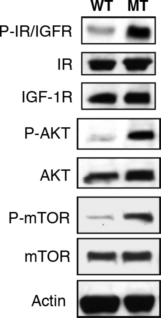
Activation of Akt and IGF1R/IR in Ankrd26 MT mouse heart. Cardiac tissue lysate from 8-week-old Ankrd26 MT and WT mice were separated by SDS/PAGE and analyzed by Western blotting with anti-P-Akt (473), anti-Akt, anti-P-IGF1R (Tyr-1135/1136)/IR (Tyr-1150/1151), anti-P-mTOR (Ser-2448), anti-mTOR, and anti-IGF1R and anti-IR, respectively.