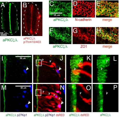
Fig. 1.
Localization of aPKCζ/λ in neuroepithelial cells of embryonic chicken spinal cord. (A and B) Cross-sections through E3 chicken neural tube revealing aPKCζ/λ and active aPKCζ/λ localization at ×10 magnification. (C–H) Open-book preparation of E3 neural tube was stained with the indicated antibodies and visualized at ×63 magnification. (E and H) Merged images show overlapping distribution of aPKCζ/λ and cell-adhesion proteins. (I–P) Chicken embryos were microinjected and electroporated at E2 with low amounts of dsRED expression vector to trace the radial processes of individual progenitors in few cells. Embryos were fixed at E3 and E4 and stained with aPKCζ/λ and p27Kip1 antibodies. (I, J, M, and N) Twenty optical sections through the z axis, 20 nm each, were collected on a Zeiss laser confocal microscope by using a ×63 objective, and this Z series was stacked by using PASCAL software. Nuclear-localized p27Kip1 is indicated with solid arrowheads. (J and N) An individual p27Kip1− progenitor (J) and a p27Kip1+ cell (N) are traced with open arrowheads. (K and L) The area indicated by the dotted box in J is magnified to show the end feet of two dsRED-labeled progenitors (open arrowheads). (O and P) Similar magnification of the area indicated by the dotted box in N. The process of the p27Kip1+ cell shown within the boxed area in N is magnified in O and P and traced with open arrowheads. The stacked Z series were rotated for the images in K, L, O, and P.

