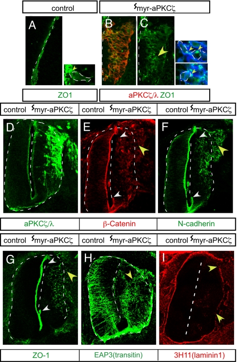
Fig. 3.
Redistribution and loss of apical adherens junction proteins and disruption of basal lamina after myr-aPKCζ expression. (A) Normal apical localization of ZO-1 at the apical, luminal margin. (B–I) Ectopic localization of adherens junction proteins at E3 (B and C) and E4 (E–G) or disruption of apical–basal polarity in radial glial processes at E4 (EAP3 antitransitin intermediate filament antibody staining) (H) and basal lamina at E4 (chicken laminin 1 antibody staining) (I) after introduction of myr-aPKCζ (B–I) at E2. Abnormal distribution is indicated with yellow arrowheads. (E–G) The region of the midline showing loss of apical junction proteins is indicated within white arrowheads.

