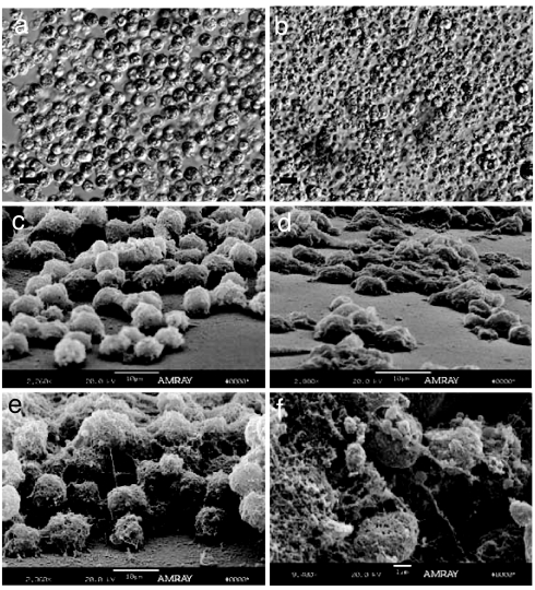
Fig. 1.
Relief contrast and SEM images of coral cell cultures. (a and b) Relief contrast of X. elongata (a) and M. digitata (b). (c and d) SEM images showing the different types of ECM monolayer of adherent cells from X. elongata (c) and M. digitata (d). (e and f) multilayer adherent cell aggregates from X. elongata (e) and nonadherent cell aggregates from M. digitata (f). [Scale bars, 10 μm (a–e) and 1 μm (f).]

