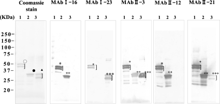
FIG. 3.
Western blot detection of MAb reactivity to SARS CoV N protein. (Coomassie stain panel) Coomassie blue staining of purified recombinant SARS CoV N protein. The recombinant viral protein was loaded at 1.0 μg/lane and analyzed on a 5 to 20% SDS-PAGE and stained by Coomassie blue dye. (MAb I-16, MAb I-23, MAb II-3, MAb II-12, and MAb II-21 panels) Western blot detection of recombinant SARS CoV N protein by MAbs. Recombinant N proteins were transferred to polyvinylidene difluoride membrane after SDS-PAGE, and each membrane was reacted to different MAbs (I-16, I-23, II-3, II-12, and II-21). Lane 1, full-length N protein was applied; lane 2, truncated N protein (NΔ283-422) was applied; and lane 3, truncated N protein of SARS CoV (NΔ1-141) was applied. ○, completely stained and partially degraded N protein (full length); •, completely stained N protein ΔN283-422; ▴, completely stained and partially degraded N protein (NΔ1-141); *, anti-N MAb reacted to complete and partially degraded N protein (full length); **, anti-N protein MAb reacted to complete N protein (NΔ283-422); ***, anti-N protein MAb reacted to complete and partially degraded N protein (NΔ1-141).

