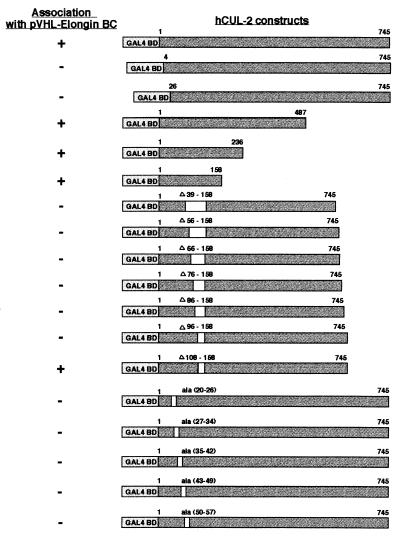
Figure 3.
Mapping the pVHL binding region on hCUL-2. Various pAS2–1-hCUL-2 constructs were cotransformed with pACT1-VHL into CG1945-BC yeast, and growth on SD + CSM−Ade−Leu−Trp−His and β-gal activity were measured. Amino acid residues are indicated on top of every hCUL-2 construct, as well as residues deleted or replaced by alanines. Expression of proteins was verified by Western blot analysis (data not shown).

