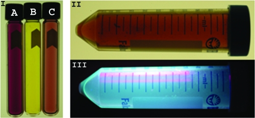
FIG. 4.
Supernatants from cryptococcal cultures demonstrate coloration, and pigmented cells extracted with methanol were fluorescent. Tube B (section I) is the amber color of the filtered supernatant from m-DTDP grown C. gattii cultures. Tube A (section I) shows the pink/fuchsia color resulting from addition of the Salkowski reagent to the supernatant. Tube C (section I) shows the lighter pink color resulting from addition of hydrochloric acid, nitric acid, or phosphoric acid to the supernatant. The methanol-extracted brown pigments from m-DTDP cells are shown in section II. The methanol-extracted pigments and chemical compounds were highly fluorescent at 365 nm (section III).

