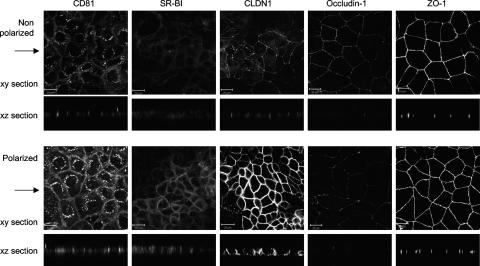
FIG. 4.
HCV receptor and TJ protein localization. Caco-2 cells were seeded onto glass coverslips under conditions optimized to achieve nonpolarized and polarized monolayers, as detailed in the legend to Fig. 1. Cells were stained with antibodies specific for CD81, SR-BI, CLDN1, occludin 1, and ZO-1. Bound antibodies were visualized with an anti-mouse-Alexa 488 secondary antibody and confocal microscopy. The large panel represents x-y sections, and the small panel represents x-z sections taken from each of the corresponding x-y sections, with the arrow indicating the plane the z section was taken from. z sections were compiled by taking 0.76-μm steps through each x-y section. The scale bars represent 20 μm.

