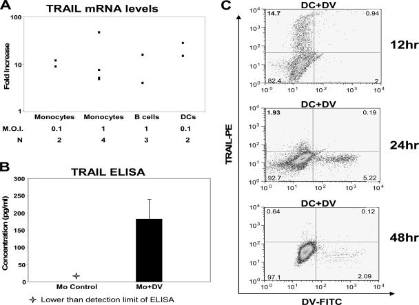
FIG. 2.
TRAIL mRNA and protein induction in DV-infected cells. (A) TRAIL mRNA levels were measured by qRT-PCR. Monocytes were infected with DV at an MOI of 0.1 and 1 PFU/ml, DCs were infected with DV at an MOI of 0.1 PFU/ml, and B cells were infected with DV at an MOI of 1 PFU/ml for 48 h. TRAIL mRNA expression was quantified by qRT-PCR analysis on total RNA extracts. β-Actin mRNA, a constitutively expressed protein, was used as a control probe. Data shown are representative of multiple (N) experiments. (B) TRAIL protein levels were measured in cell lysates. Monocytes (Mo) were infected with DV at an MOI of 1 PFU/cell and then cultured for 48 h. Levels of TRAIL protein were quantified in cell lysates using a TRAIL ELISA (R&D Systems). Data shown are representative of three experiments. (C) Intracellular TRAIL protein levels were determined in DCs infected with DV for 12, 24, and 48 h at an MOI of 0.1. Cells were treated with brefeldin A for the last 8 h of each time point. Levels of TRAIL protein were quantified by flow cytometry using TRAIL-PE (BD Biosciences). Data shown are a representation of two experiments. FITC, fluorescein isothiocyanate.

