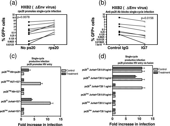
FIG. 9.
ps20 promotes HIV entry via fusion. (a) Cells (2 × 10⁵) from NP clones (86 1-1 and 8.5.7.05) were precultured for 18 h with 1 μM rps20 and then infected with replication-defective GFP-encoding lentivirus particles pseudotyped with HIV-1 envelope (strain HXB2); three doses were offered (dose 1, 0.1 to 0.5% GFP+ cells; dose 2, 0.5 to 1% GFP+ cells; dose 3, >2% GFP+ cells). Cultures were maintained in 30 IU/ml IL-2. Mean percentages of GFP+ cells at 6 days postinfection in the absence and presence of rps20 are shown. Paired t testing was used to calculate statistical differences between treatments. (b) Cells (2 × 10⁵) from two P clones (86 1-3 and 8.16.7.05) were precultured for 18 h with 5 μg/ml control mouse IgG or IG7 and then infected with replication-defective GFP-encoding lentivirus particles exactly as described for panel a. Percentages of GFP+ cells for each culture at 6 days postinfection in the presence of control IgG versus that of IG7 are shown. Paired t testing was used to calculate statistical differences between treatments. (c and d) Cells (2 × 10⁵) were infected with NL4-3 (10 ng p24-CA/million cells) in the presence (treatment) or absence (control) of the following modulators: fusion inhibitor T20 (kind gift of B. Peters) and 0.03 μg/ml rps20. Where treatment included culturing with 5 μg/ml IG7, the control was 5 μg/ml normal mouse IgG. Twenty-four hours later, cells were washed and cell lysates harvested for p24-CA measurement. Increases (n-fold) in the infection of each sample were based on an additional sample treated in parallel, exposed to HIV for 1 min, and taken as 1.0.

