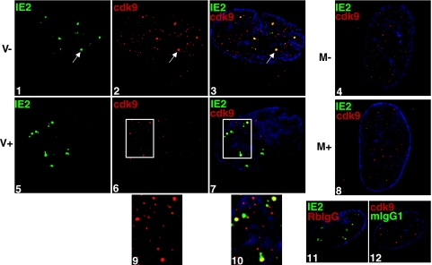
FIG. 2.
Treatment with roscovitine impairs accumulation of cdk9 at the sites of viral IE transcription. G0-synchronized cells were released into G1, infected with HCMV Towne at an MOI of 5 or with mock supernatant, and seeded onto glass coverslips. Cells were treated with 16 μM roscovitine or DMSO at the time of infection. At 8 h p.i., cells were washed with PBS, fixed in formaldehyde, permeabilized, and immunostained with IE2 (IgG1) and cdk9. Specific antibody staining was detected with fluorescein isothiocyanate- or Cy3-conjugated isotype-specific secondary antibodies. For controls, one of the specific antibodies of the pair was replaced with an isotype-specific normal IgG. Nuclei were stained with Hoechst dye. The white arrow in each panel indicates a region of colocalization, although more are present. The white boxes in panels 6 and 7 are enlarged in panels 9 and 10, respectively, with the modification of a 200% magnification and a 200% Cy3 intensity using Adobe Photoshop v. 7.0. All of the images are confocal optical sections of 0.2 μm at a magnification of ×1,000 under oil immersion. M, mock treated; V, HCMV infected; −, DMSO treated; +, roscovitine treated at the time of infection.

