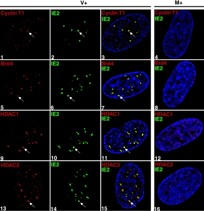
FIG. 5.
Cyclin T1, Brd4, HDAC1, and HDAC2 are able to localize to the sites of viral IE transcription during infection in the presence of roscovitine. G0-synchronized cells were released into G1, infected with HCMV Towne at an MOI of 5 (panels 1 to 3, 5 to 7, and 13 to 15) or with mock supernatant (panels 4, 8, 12, and 16), and seeded onto glass coverslips. Cells were treated with 16 μM roscovitine or DMSO (see Fig. 1) at the time of infection. Only the roscovitine-treated samples are shown in this figure. At 8 h p.i., cells were washed with PBS, fixed in formaldehyde, permeabilized, and immunostained with an antibody specific for the protein indicated in each panel. Specific antibody staining was detected with fluorescein isothiocyanate- or Cy3-conjugated isotype-specific secondary antibodies. For controls, one of the specific antibodies of the pair was replaced with an isotype-specific normal IgG (not shown). Nuclei were stained with Hoechst dye. The white arrow in each panel indicates a region of colocalization, although more are present. All of the images are confocal optical sections of 0.2 μm at a magnification of ×1,000 magnification under oil immersion. M, mock treated; V, HCMV infected; +, roscovitine treated at the time of infection.

