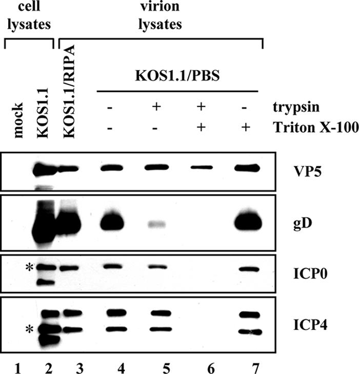
FIG. 4.

ICP0 and ICP4 are localized to the tegument layer of HSV-1 virions. Vero cells were infected with WT HSV-1, and extracellular virions were purified on a Ficoll gradient as described in Materials and Methods. Virions were either solubilized in RIPA buffer or resuspended in PBS. Those resuspended in PBS were then treated with trypsin (0.1 mg/ml) in either the presence or absence of 1% Triton X-100 for 20 min at 37°C. Equivalent amounts of cell lysates as well as virion samples were analyzed by immunoblotting for the viral proteins indicated. For ICP0 and ICP4, asterisks mark the positions of the major 110-kDa and 175-kDa species, respectively.
