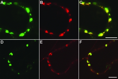
FIG. 1.
Confocal laser-scanning microscopy of N. benthamiana epidermal cells transiently expressing fluorescent fusion proteins after agroinfiltration. (A to C) Colocalization of 30K-GFP (A) and DsRed-18K (B). (C) Superposition of the images in panels A and B. (D to F) Colocalization of Hsp70h-RFP (E) with GFP-18K (D). (F) Superposition of the images in panels D and E. Scale bars = 5 μm.

