
FIG. 9.
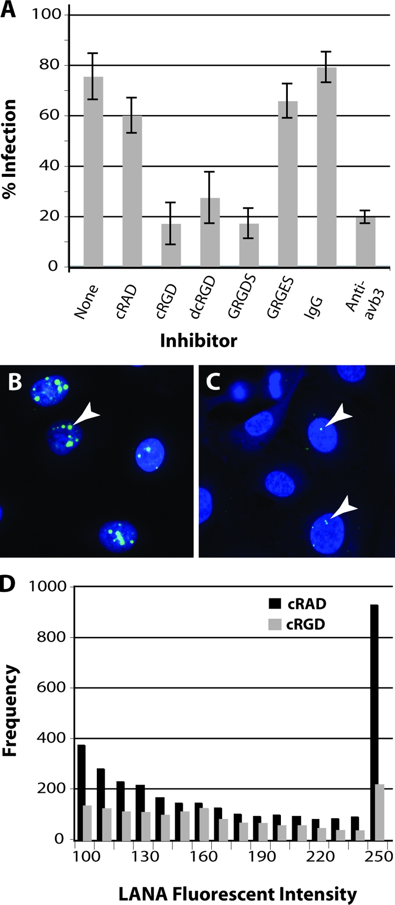
KSHV infection is inhibited by αVβ3-specific ligands and function-blocking antibodies. Infection-blocking studies were performed by pretreating HT1080 cells (4°C) with RGD-containing peptide ligands, including the linear GRGDS peptide and the αVβ3-specific cyclic (cRGD) and dicyclic (dcRGD) peptides or the αVβ3 function-blocking antibody (LM609). Controls included the linear GRGES and cyclic cRAD peptides and normal mouse IgG. Gradient-purified KSHV virions titrated to yield an infection in 75% of the cells were incubated with the cells for 1 h (4°C) in the presence of peptide or antibody. Unbound KSHV was removed by washing, and the percentage of cells expressing KSHV LANA was determined 24 h later in multiple fields by confocal microscopy (A). The standard deviation is shown. Representative micrographs showing nuclear LANA expression in control cRAD-treated (B) and cRGD-treated (C) HT1080 cells infected with KSHV are shown. Arrows indicate representative dots of LANA fluorescence. LANA fluorescence was quantitated, and the frequency and intensity (range, 100 to 255) of fluorescent pixels within LANA dots in individual nuclei (n = 30) were plotted (D).