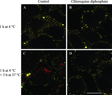
FIG. 4.
Effects of CQ treatment on PCV2 attachment and internalization. PCV2 VLP were either allowed to bind to PK-15 cells for 1 h at 4°C or further incubated for 3 h at 37°C in the absence or presence of 125 μM CQ and then fixed with 3% paraformaldehyde. PCV2 VLP were stained with F190 monoclonal antibody and FITC-conjugated goat anti-mouse, followed by permeabilization of the cells. PCV2 VLP were stained again with F190 and Texas Red-conjugated goat anti-mouse antibodies. (A to D) Bound PCV2 VLP showed both green and red fluorescence (yellow), while internalized PCV2 VLP showed only red fluorescence in merged confocal images of single z sections. Internalized PCV2 VLP were visible in untreated cells (C), while no PCV2 VLP were visible within the cell in CQ-treated cells (D), most probably due to increased disassembly of internalized PCV2 VLP. Bar, 20 μm.

