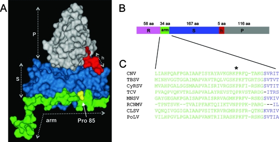
FIG. 1.
(A)Tertiary structure of the CNV CP C subunit with ordered arm, S-, and P-domains, with the hinge (h) indicated. The structure is based on homology modeling using the X-ray crystal structure of the TBSV CP subunit (27). The location of the conserved Pro85 residue is also shown. The disordered R-domain is not shown. (B) The CP linear structure is shown, using the same color scheme as in panel A. The number of amino acids in each domain is given. (C) Alignment of the arm region and the junction at the S-domain of several members of the Tombusviridae family, demonstrating conservation of the Pro residue (Pro85 in CNV) (asterisk) in this region. CNV, TBSV, and Cymbidium ringspot virus (CyRSV) are tombusviruses, TCV and Melon necrotic spot virus (MNSV) are carmoviruses, Red clover necrotic mosaic virus (RCNMV) is a dianthovirus, and CLSV (Cucumber leaf spot virus) and PoLV (Pothos latent virus) are aureusviruses.

