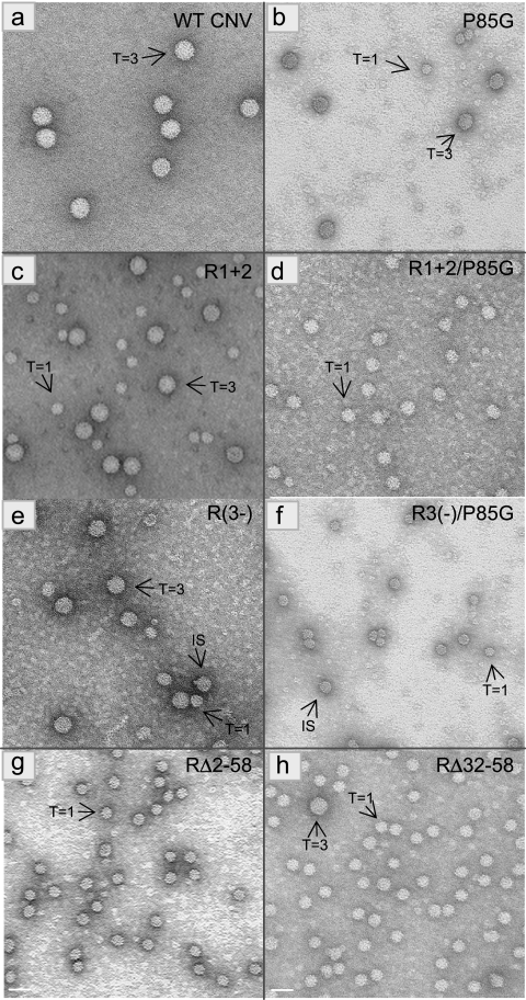
FIG. 5.
TEM of particles extracted from leaves infected with the indicated mutants. Particles were stained with 2% uranyl acetate and photographed at a magnification of ×80,000. The T=3 or T=1 morphology of particles is shown with arrows. The bar corresponds to approximately 34 nm. Table 1 contains a summary of the various particle types observed for each mutant.

