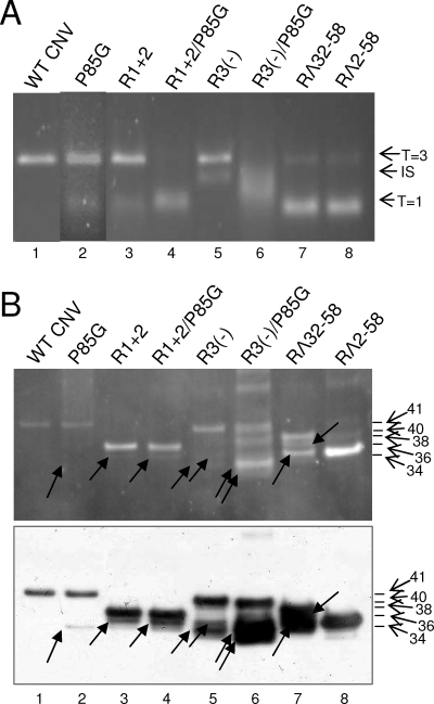
FIG. 6.
Analysis of particles obtained from plants infected with single and double P85G and R-domain mutants. (A) Approximately 1.5-μg portions of purified virions were electrophoresed through a 2% TB-agarose gel. The gel was stained with SybrSafe (Invitrogen). The arrows on the right indicate possible particle morphologies as assessed by TEM (Fig. 5). (B) Virions of the indicated mutants were denatured with 1× LDS buffer, electrophoresed through 4 to 12% NuPage-MES (morpholineethanesulfonic) gels, and blotted to PVDF membranes. Membranes were then treated with SYPRO-RUBY blot stain and visualized by epifluorescence (top). The blot (bottom) was probed using a mixture of three CNV antibodies known to be specific to the arm and the R- and S-domains (18) (unpublished data). The mass of WT CNV CP and the predicted molecular masses of the CPs of the mutants are indicated by the arrows and summarized in Table 1. Arrows within the blot point to the major truncated proteins detected in each mutant (Table 1). Standards (not shown) were SeeBlue+2 and Mark 12 (Invitrogen).

