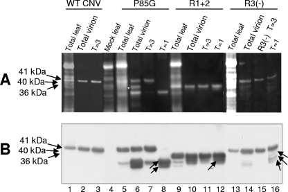
FIG. 8.
Denaturing polyacrylamide gel electrophoresis of protein extracted from infected leaves (TL), total virions (V), and gel-purified particles of P85G, R1+2 and R3(−). WT CNV-, P85G-, R1+2-, and R3(−)-infected leaves were ground in liquid nitrogen. A portion of the ground material was used to extract total protein and another portion was used for total virion extraction. Virions were electrophoresed through a 2% agarose-TB gel, and particles corresponding to T=3, IS, and T=1 particles were purified from gel slices. (A) Samples were denatured with 1× LDS buffer, electrophoresed through 4 to 12% NuPage-MES gels, and blotted to PVDF membranes. Membranes were then treated with SYPRO-RUBY blot stain and visualized by epifluorescence. (B) Western analysis of the blot shown in panel A using a mixture of CNV antibodies specific to the R-, arm, and S-domains. The predicted sizes of CNV CP (41 kDa) as well as those of full-length P85G (41 kDa), R1+2 (36 kDa), and R3(−) (40 kDa) are shown on the left. Arrows within the blot point to truncated proteins found within T=1 virions (Table 1). The white asterisk indicates the position of an unidentified nonviral protein that copurified with P85G particles. Size standards (not shown) were SeeBlue+2 and Mark 12 (Invitrogen).

