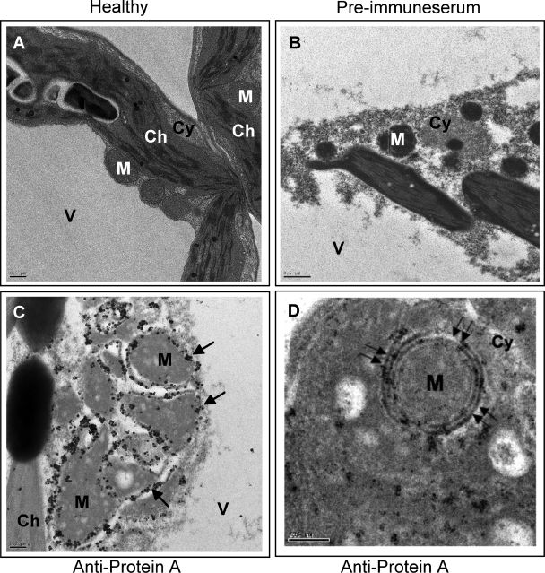
FIG. 3.
Immunogold EM localization of FHV protein A. (A) EM of a section of healthy N. benthamiana leaf showing vacuole (V), chloroplast (Ch), cytoplasm (Cy), and mitochondria (M). (B) EM of a section of F1-infiltrated N. benthamiana leaf treated with preimmune serum. (C) EM showing localization of FHV protein A on mitochondria of N. benthamiana infiltrated with the F1 agrotransformant. Arrows show the location of gold particles on outer mitochondrial membranes. (D) EM (at a higher magnification) reveals localization of gold particles to outer double-walled mitochondrial membrane (double arrows). Scale bars = 0.5 μm (panels A and B), 0.2 μm (panel C), and 200 nm (panel D).

