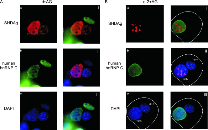
FIG. 3.
A heterokaryon assay demonstrated that subcellular localization of the SHDAg-NoLS mutant was confined to the nucleolus. Huh7 cells seeded on coverslips were cotransfected with 3 μg of pCDm2AG as well as 1 μg of the expression plasmid of SHDAg (d+AG) or clone d-2 (d-2+AG). The transfected cells were incubated and then fused with NIH 3T3 cells (see Materials and Methods). Five hours after heterokaryon formation, cells were fixed and analyzed by immunofluorescence microscopy. Frames I to III in panels A and B are merged images of the immunofluorescence results as follows: frames I, SHDAg and human hnRNP C (C1/C2); frames II, SHDAg and DAPI (4′,6′-diamidino-2-phenylindole) staining; and frames III, human hnRNP C (C1/C2) and DAPI staining. White lines mark cell boundaries.

