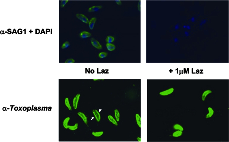
FIG. 5.

Treatment with Laz blocks anti-SAG1 binding. Tachyzoites were used in an immunofluorescence assay to detect SAG1, which normally stains the outer membrane of the parasite (green, top left panel). When tachyzoites were treated with 1.0 μM Laz, anti-SAG1 is no longer able to bind the parasite surface (top right panel). 4′,6-Diamidino-2-phenylindole (DAPI), which binds DNA, was used as a costain for reference (blue). The lower panels show staining with antibody generated against Toxoplasma (anti-Toxoplasma) with (right) or without (left) the 1.0 μM Laz treatment. Arrows highlight the more-intense staining of the parasite plasma membrane (seen as a ring encircling the parasite), to be contrasted with the less-intense membrane staining (lack of ring) in the Laz-treated parasites.
