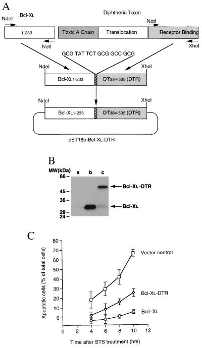
Figure 1.
Construction, Western blotting, and bioactivity of transfected Bcl-xL–DTR and Bcl-xL. (A) Schematic diagram of the chimera, Bcl-xL–DTR. The fusion gene, Bcl-xL–DTR, was inserted into the vector, pET16b yielding a histidine tag sequence at the N terminus of the Bcl-xL–DTR gene. An 18-bp linker between the hBcl-xL and DTR genes was introduced via the PCR primers (→). (B) Western blotting of the lysates of HeLa cells transiently transfected with Bcl-xL and Bcl-xL–DTR. At 20 hr after transfection with Bcl-xL–DTR or Bcl-xL genes, 106 HeLa cells were lysed in 1 ml of buffer containing 100 μg/ml leupeptin and centrifuged, and 15 μl of the supernatant was loaded onto SDS/PAGE gels, immunoblotted with anti-Bcl-xL antibody (2H12), and developed with enhanced chemiluminescence. Lane a, untransfected cells; lane b, cells transfected with Bcl-xL; lane c, cells transfected with Bcl-xL–DTR. A small amount of endogeneous Bcl-xL is present in lanes a and c. (C) Transient transfection of Bcl-xL (○) and Bcl-xL–DTR (⋄) genes into HeLa cells shows an inhibition of cell death induced by the addition of 0.8 μM STS compared with pcDNA3 vector transfected cells (□).

