Figure 3.
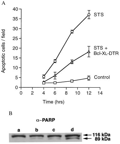
Inhibition of apoptosis. (A) The time course of apoptosis induced by STS in Cos-7 cells with and without Bcl-xL–DTR protein. Cos-7 cells at 3 × 10⁴ cells per cm2 in 100 μl of DMEM with 10% FBS were incubated with 0.1 μM STS (○), 0.1 μM STS plus 4.8 μM Bcl-xL–DTR protein added to the medium (▵), or 20 μl of PBS (□). Apoptotic cells were quantified by staining with Hoechst dye no. 33342. Results are presented as the average number of cells per field (magnification ×160). For each point, at least five fields were counted in each of at least three wells. Bcl-xL–DTR dramatically decreased the rate of apoptosis in Cos-7 cells. Six different batches of Bcl-xL–DTR were found to have activity, and the apoptosis prevention activity was stable for at least 5 months when Bcl-xL–DTR was stored at 4°C. (B) Prevention of PARP cleavage by Bcl-xL–DTR. HeLa cells were treated with two different batches of Bcl-xL–DTR at 1.48 μM or 1 μM. Fifteen hours later, cells were treated again with Bcl-xL–DTR at 1.48 μM or 1 μM. Immediately after the second treatment, 0.8 μM STS was added. Three hours later, cell lysates were made and loaded onto SDS/PAGE gels, immunoblotted with anti-PARP polyclonal antibody, and developed with enhanced chemiluminescence. Lane a, HeLa cells not incubated with STS; lane b, HeLa cells treated with STS plus 1 μM Bcl-xL–DTR protein; lane c, HeLa cells treated with STS plus 1.48 μM Bcl-xL–DTR protein; lane d, HeLa cells treated with STS.

