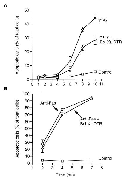
Figure 4.
Bcl-xL–DTR inhibition of apoptosis induced by γ-radiation and α-Fas antibody. At various times after induction of apoptosis by γ-radiation or α-Fas antibody, viable and apoptotic cells were counted by using Hoechst dye no. 33342. (A) Jurkat cells were plated at 105 cells per ml in serum-free RPMI-1640 medium with insulin and transferrin and γ-irradiated at 10 grays a few minutes after addition of Bcl-xL–DTR to a concentration of 4.68 μM. Control cells were not irradiated and not treated with Bcl-xL–DTR. (B) Jurkat cells were plated at 105 cells per ml in serum-free RPMI-1640 medium with insulin and transferrin, and treated with 100 ng/ml anti-Fas antibody (CH11, Upstate Biotechnology, Lake Placid, NY) minutes after addition of Bcl-xL–DTR to a concentration 4.68 μM. In contrast to irradiation-induced apoptosis of Jurkat cells, Bcl-xL–DTR had little inhibitory effect on apoptosis induced by anti-Fas antibody. Control cells were treated with PBS and no anti-Fas antibody.

