
Figure 3.
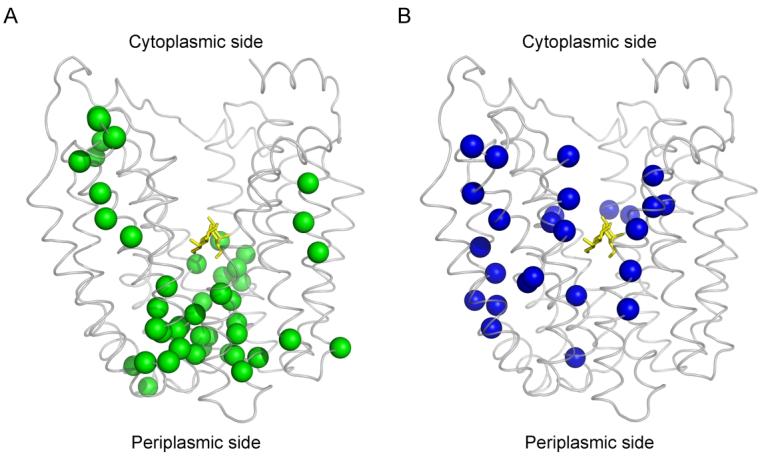
Distribution of single-Cys replacements exhibiting TDG-induced changes in TMRM reactivity. Seventy-one single-Cys LacY mutants were labeled with TMRM in the absence and presence of TDG as described in Fig. 2. Positions of Cys replacements that exhibit changes in TMRM reactivity are superimposed on the backbone of LacY (Protein Data Bank ID code 1PV7; www.pdb.org). LacY is viewed perpendicular to the membrane with the N-terminal helix bundle on the left and the C-terminal bundle on the right. (A) green spheres, increased TMRM reactivity at positions 2, 3, 8, 12, 14, 17, 24, 25, 28, 29, 30, 31, 32, 42, 44, 45, 49, 53, 70, 71, 96, 100, 136, 157, 158, 159, 160, 161, 241, 242, 244, 245, 246, 248, 265, 291, 295, 298, 308, 315, 359, 361, 362, 363 and 364; (B) blue spheres, decreased TMRM reactivity at positions 4, 5, 11, 15, 21, 22, 27, 34, 60, 81, 84, 86, 87, 88, 122, 141, 145, 148, 264, 268, 272, 327, 329, 331, 356 and 357. TDG is shown as a stick model at the apex of the inward-facing cavity (yellow sticks).