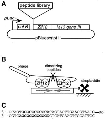
Figure 1.
(A) Sketch showing key segments of the phagemid. (B) Expected arrangement of fusion proteins at the target DNA. Phage displaying two copies of a dimerizing peptide–Zif12 fusion can form stable complexes with the biotinylated target DNA site, which contains an inverted repeat of the Zif12-binding site. The phage–DNA complexes are captured by streptavidin coupled to a solid support, and phage that bind less tightly are washed away. (C) The DNA site used for affinity selection of phage, with the two juxtaposed Zif12-binding sites in bold.

