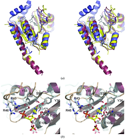
Figure 4.
(a) Ribbon diagram showing the superimposition of the SaGMK open state (coloured yellow), closed state (coloured raspberry) and the mouse GMK closed state (coloured blue). The SaGMK ligands, sulfate and GMP, are in grey. (b) Stereo diagram showing the superimposition of the open conformation of the SaGMK active site (transparent representation) onto the closed conformation. The GMP and sulfate are in standard atom colours; the potassium ion is coloured cyan.

