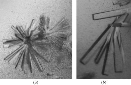
Figure 2.
(a) A cluster of rod-shaped crystals of the AtCSN7 core as obtained in the initial screen. The cystals are approximately 0.05–0.1 mm in length. (b) Rod-shaped crystals of the AtCSN7 core obtained after lowering the magnesium acetate concentration. The crystals are approximately 0.15–0.3 mm in the longest dimension. The crystallization conditions for these crystals were a protein concentration of 10 mg ml−1 with a reservoir comprising 23%(w/v) PEG 8000, 0.1 M sodium cacodylate pH 7.2 and 0.08 M magnesium acetate.

