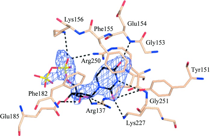
Figure 3.
UDP-binding site and the omit difference electron-density map. The map has coefficients |F o − F c|, αc and is contoured at 3.5σ. F o represents the observed structure factors, F c the calculated structure factors and αc the calculated phases from which ligand contributions were omitted. Protein residues within 4 Å of UDP are labelled and putative hydrogen bonds (within 3.6 Å) are shown as black dashed lines. Atoms are coloured as described in Fig. 2 ▶, with the Cα atoms of the protein coloured salmon.

