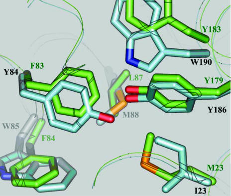
Figure 2.
An overlay of the PmSOD1 (green) and PmSOD2 (blue) structures reveals key mutations in a highly conserved triad (Phe-Try-Trp) of Fe-SOD. PmSOD1 has the unusual sequence Phe-Phe-Phe. The Tyr84 to Phe83 change is structurally compensated by the Ile23 to Met23 change owing to the larger volume of Met relative to Ile. Overall, PmSOD1 displays a more hydrophobic second shell of amino-acid residues than PmSOD2 around the metal site, i.e. Phe83, Phe84 and Tyr183.

