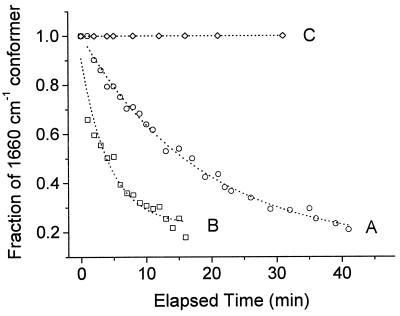
Figure 2.
Effect of quinol on the rate of conformational change in heme a3. The decrease in the heme a3 formyl line at 1,660 cm−1 was monitored as a function of time after reduction, in the presence and in the absence of quinol, at two different pH values. The enzyme was reduced anaerobically by dithionite at pH 6, 100 mM Mes, in the absence (A) and in the presence (B) of 1.6 mg/ml decylubiquinol. At pH 2.6 (C), the population of the 1,660-cm−1 conformer remained invariant in both the presence and the absence of decylubiquinol. The relative population of the 1,660-cm−1 (to that of 1,667 cm−1) conformer was calculated by curve fitting using two Lorentzians.

