
FIG. 3.
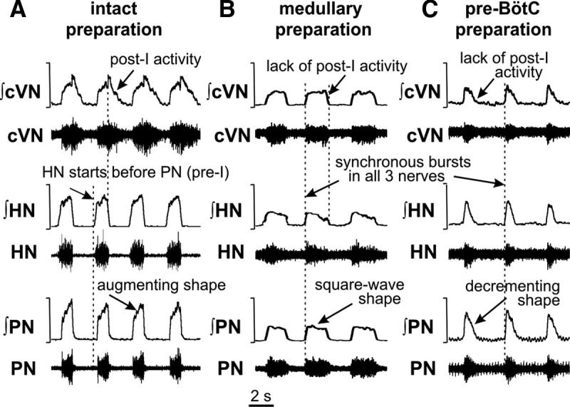
Representative activity patterns of phrenic (PN), hypoglossal (HN), and central vagus (cVN) nerves recorded from intact (A), medullary (B), and pre-BötC (C) preparations. Each panel shows raw (bottom traces) and integrated (top traces) motor nerve discharge. Vertical dashed lines in A indicate onsets of HN inspiratory burst and the postinspiratory component of cVN activity; dashed lines in B and C indicate synchronous onset of inspiratory bursts in all nerves.