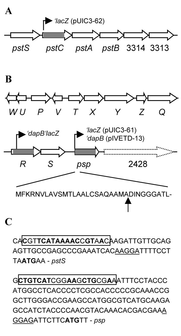
Figure 1.
Genetic organization of the pst (A) and psp (B) loci in P. fluorescens SBW25. Regions deleted in mutants PBR827 (ΔpstC or Δpflu3317), PBR828 (ΔhxcR or Δpflu2424) and PBR826 (Δpsp or Δpflu2427) are marked by grey. Positions of the promoterless 'lacZ and 'dapB fusions are shown by arrowed lines. The predicted signal peptide cleavage site is indicated by vertical arrow. ORF Pflu2428 is not drawn to scale and shown by discontinued arrow bar. (C) Promoter regions of pst and psp operons. The putative translational start and ribosome-binding sites are indicated by bold type and underlined letters, respectively. The predicted Pho box sequences are boxed and nucleotides identified in the E. coli Pho box consensus [CTGTCATA(AT)A(TA)CTGT(CA)A(CT)] [7] are highlighted by bold type.

