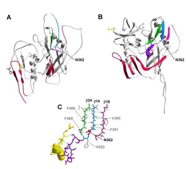
Figure 7.
Structural modelling of N362. Structures of the unliganded SIV gp120 (A) and CD4-bound JR-FL gp120 (B). The β-14 (cyan), β-18 (pink) and β-24 strands (green) are highlighted. The CD4 binding loop is highlighted in purple. Asn362 (Thr378 in SIV) is labelled (cyan). Elements of the bridging sheet are highlighted in red. Potential hydrogen bond donors for N362 within the β-14, β-18 and β-24 strands are shown in (C) and are colored as in (A) and (B). CD4 residues contacting the CD4bs of gp120 are colored in yellow, and the molecular surface of Phe43 of CD4 is shown to illustrate the "Phe43 pocket" of the gp120 binding site of CD4. N362 is labelled and highlighted in red. Putative hydrogen bond partners are labelled in grey. Hydrogen bonds are depicted as dotted green lines. For simplicity, only the N362 hydrogen bond with R465 is shown.

