
Fig. 5. Activation of either mAChRs or mGluRs facilitates DSE at the MCF-GC synapse.
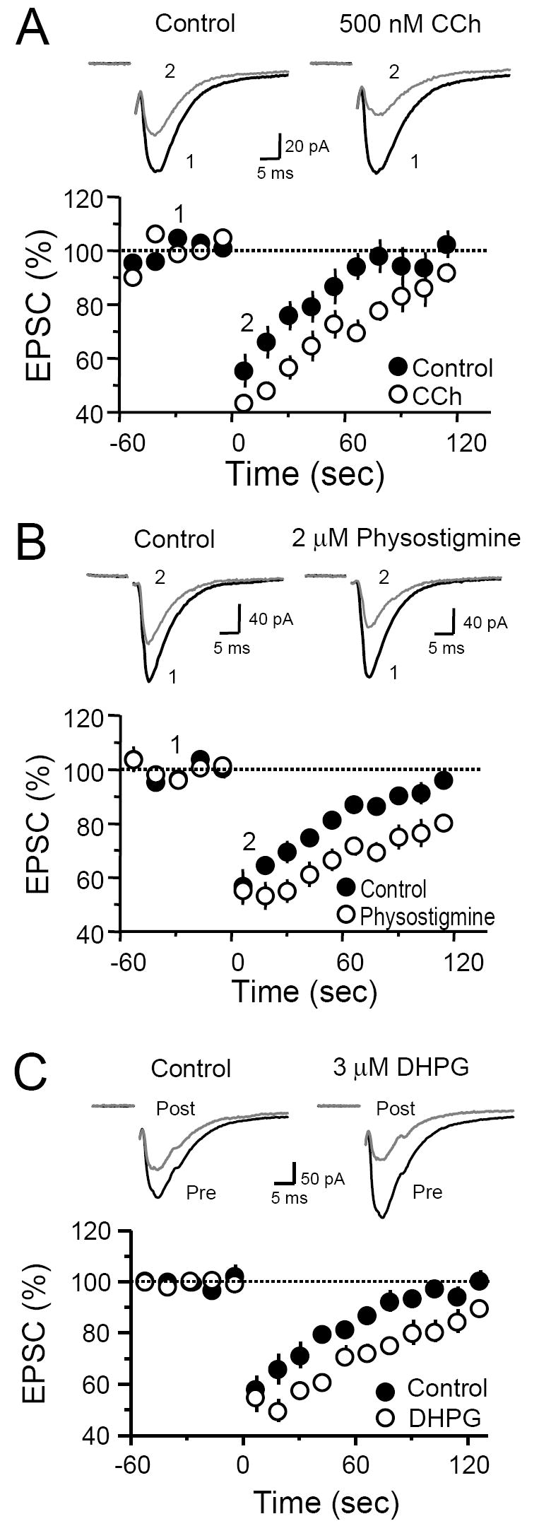
(A-C) Time course plots of DSE before and after bath application of 500 nM CCh (A), 2 μM physostigmine (B) and 3 μM DHPG (C). Top of each panel (A-C), representative EPSC traces from single experiments.